PLANKTON
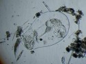
Plankton can be examined very easily, so for amateurs it is wise to start prasticing microscopy this way, but even examining plankton requires some refinement: The proverbial "Miracle of Life in one Drop of Water" will usually be a great disappointment because of the low concentration of plankton organisms. You need a professional plankton net, made of fine gauze with a mesh size of about 0.05 - 0.02 mm. This net is pulled through the water several times or else you may filter some ten litres of water through such a net using a bucket. Samples thus collected should be kept cool and must be investigated as soon as possible. The plentiful amount of different forms is always overwhelming! Afterwards the sample is preserved by mixing 10 ml sample with 1 ml formaldehyde, but it is impossible to stabilize all types of organisms - only algae and animals with some kind of carapace will keep their shape. It is recommended to add some drops of glycerol to prevent desiccation and a small crystal of copper acetate to stabilize chlorophyll. More detailed information will be found on the <tips for beginners and advanced> page.
Not all species on display are planktonic, but they will always occur in samples caught with a plankton net.
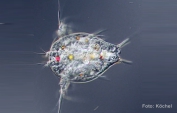
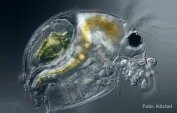
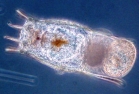
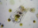
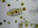
Small Crustacea and their larvae are always present in plankton samples.
|
|
|||
| Naupliuslarve | Bosmina |
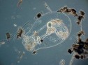
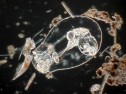
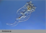
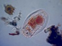
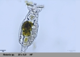
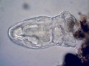
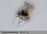
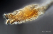
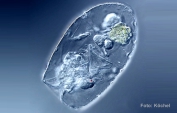
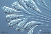
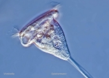
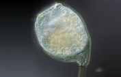
It is particularly impressive to watch Rotifers, small multicellular organisms, which are named after two special structures at their apex, which seem to whirl around like spinning wheels. Here are some pictures and clips.
 |
 |
 |
 |
 |
| Keratella quadrata | Asplanchna | Asplanchna | Asplanchna | |
 |
 |
 |
 |
 |
| Brachyonus | ||||
 |
 |
|||
| Asplanchna |
 |
 |
 |
 |
Please wait while buffering is under way! |
| Rotifer | Rotifer | Flagellates | Flagellates |
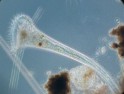
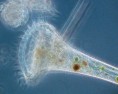
Another interesting
group are the very vivid unicellular Ciliates.
Here some examples:
 |
 |
 |
 |
 |
| Trompetentierchen | Ciliatenkolonie | Pantoffeltierchen | Glockentierchen | |
 |
||||
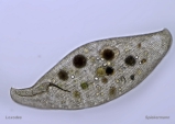
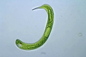
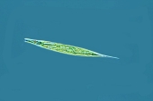
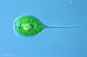
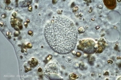
Monocellular Flagellates are common in plankton samples. This heterogeneous
group links the realm of plants with that of animals because many species cntain
chlorophyll (autotrophic organisms) while others don´t (heterotrophic
organisms).
 |
 |
 |
 |
 |
| Augentierchen | Augentierchen | Phacus | Synura uvella | Synura uvella |
 |
||||
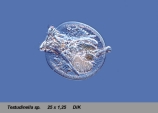
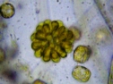
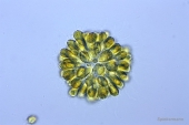
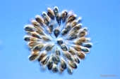
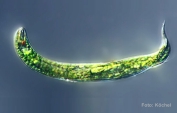
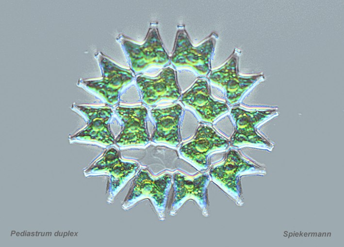
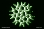
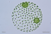
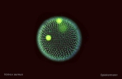
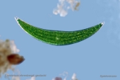
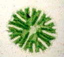
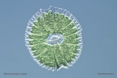
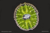
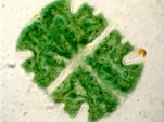
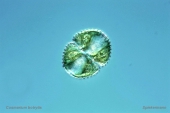
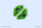
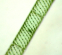
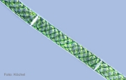
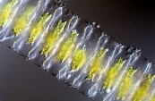
Chlorophyceae are ubiquitous in plankton samples. Some of the most beautiful algae are Desmids. Although they are not planktonic organisms, they occur very often in plankton samples. They are abundant in boggy areas and swamps of slightly acidic water. Squeeze the wet moss found in such places, sieve the water through a plankton net and you will find many different species!
 |
 |
 |
 |
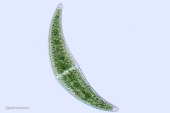 |
| Pediastrum | Pediastrum | Volvox | Volvox | Closterium |
 |
 |
 |
 |
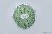 |
| Closterium | Micrasterias | Micrasterias | Micrasterias | Micrasterias |
 |
 |
 |
 |
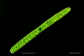 |
| Cosmarium | Cosmarium | Cosmarium | Spirogyra | Pleurotaenium |
 |
||||
| Spirogyra |
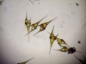
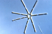
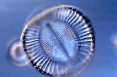
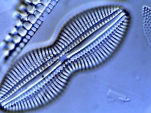
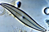
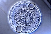
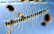
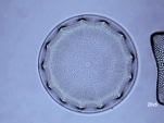
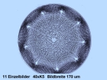
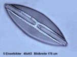
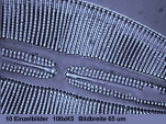
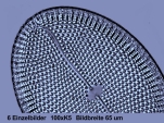
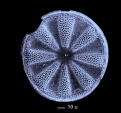
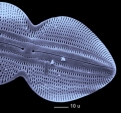
A particularly fine group are Diatoms . Planktonic species often show long setae which help them to float or to form colonies. Drops of oil in their cells enhance their buoyancy.
 |
 |
 |
 |
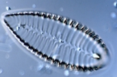 |
| Asterionella | Campylodiscus | Diploneis | Gyrosigma | Surirella |
 |
 |
 |
 |
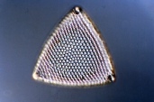 |
| Actinoptychus | Auliscus | Bellerochea | Chaetoceros | Triceratium |
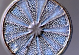
The cells of Diatoms are enclosed inskeletons of silica. These skeletons are extremely inert, so they are concentrated in the mud at the bottom of pools and lakes. To separate these skeletons a dried mud sample is heated with concentrated sulphuric acid in the open until all the organic impurities are destroyed, then the sample is washed repeatedly with water and finally immersed in alcohol. Allow a small amount of this sample to dry on the slide, then add one drop of toluene or xylene, followed by a drop of mounting medium, and after placing a coverslip on top you will possess a prepared microscopic slide. More detailed information can be found on the <hints> page.
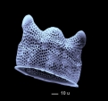
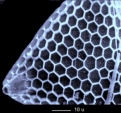
Although the structures of Diatoms can be seen with a normal microscope, they are so delicate that they can only be properly analysed when using a scanning electronic microscope ("SEM").
 |
 |
 |
 |
 |
| Aulacodiscus | Aulacodiscus | Navicula | Navicula | Mastogloia |
 |
 |
 |
 |
 |
| Biddulphia (REM) | Navicula (REM) | Triceratium (REM) | Actinoptychus (REM) | Diploneis (REM) |
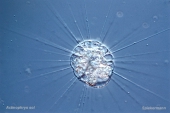
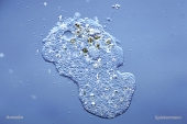
Rhizopoda are non-planktonic organisms. They inhabit the surfaces of many different substrates or they live in muddy layers. But even they are often found in plankton samples. One group (Thekamoeba) produces a carapace.
 |
 |
 |
 |
 |
| Actinophrys sol | Amoeba | Amoeba, Detail | Thekamoeba | Thekamoeba |
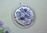
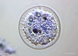
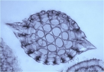
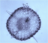
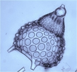
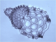
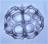
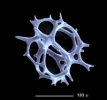
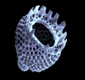
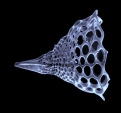
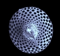
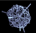
Marine monocellular Radiolarians are a related group. Alive they are rather unappealing globules of plasm, but their filigree skeletons of silica are of remarkable beauty and diversity. To see all details you must either apply "stacking" or you have to use a SEM.
 |
 |
 |
 |
 |
 |
 |
 |
 |
 |
Copyright: webmaster@mikrohamburg.de Dr.K.Spiekermann klausspiekermann@gmx.net W.Steenbock