
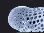
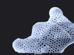
Diatoms
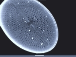
display a fascinating variety of forms and delicate structures when investigated
with a microscope, but often the spatial arrangements of structural elements
still remain enigmatic, since at higher magnifications the in this case very
restricted depth of field makes interpretation difficult. Moreover, often disturbing
image overlays with deeper structures occur, especially when inspecting diatoms
from the side ( "girdle band view").
When studying diatoms the SEM shows
its advantages, not only because of its higher resolution and depth of field,
but because the SEM is an electronic "reflected light microscope":
You see the diatom either from the outside or from the inside,
and this literally "one-sided approach" makes the analysis of the
spatial structure of the frustules an easier task. But there are also disadvantages,
because you cannot see the outer side and the inner side of the frustule at
the same time. So the internal septa remain concealed when you look at the diatom
from the outside. This makes identification difficult.
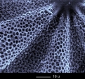
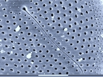
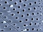
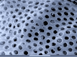
At least three different types of
shell-constructions can be distinguished, they are here referred to by the letters
A, B and C:
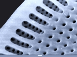
Type A: The wall of the frustules
is very thin and perforated with fine pores which allowe diffusion. Often very
small species belong to this type, such as Cyclotella and Navicula,
but also larger species like Pleurosigma and Gyrosigma.
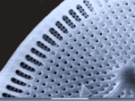
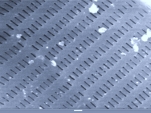
Type B: The wall of the frustules
is composed of coarse parallel or intersecting ribs, which are covered from
inside with a very thin perforated silica wall whose pores allow diffusion.
The spaces between the ribs appear as slots or areolae, the fine pores of the
inner wall usually remain invisible because they are below the maximum resolution
of the normal microscope. This is typical of many Centrales, such as
Triceratium.
Type C: Like type B, but the thin perforated silica wall is positioned outside. This is typical of many Pennales, such as Epithemia.
Probably there is one more type: Frustules with an inner and an outer perforated wall ( "sandwich structure"). It is noteworthy that HUSTEDT has described all these types more than fifty years ago!
|
|
|
|
|
|
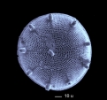
| 1 Aulacodiscus | 2 Biddulphia | 3 Navicula | 4 Diploneis | 5 Cymbella | |
|
|
|
|
|
|
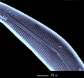
| 6 Navicula | 7 Pinnularia | 8 Coscinodiscus (?) | 9 Triceratium | 10 Triceratium | |
|
|
|
|
|
|
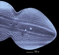
| 11 Triceratium | 12 Triceratium | 13 Triceratium, innen | 14 Triceratium, innen | 15 Arachnoidiscus, innen | |
|
|
|
|
|
|
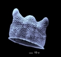
| 16 Arachnoidiscus | 17 Actinoptychus | 18 Actinoptychus | 19 Epithemia, innen | 20 Epithemia, außen |
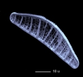
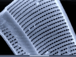
The upper row of images (Fig.1 to 5) shows species which probably all belong to type A.
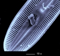
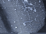
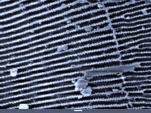
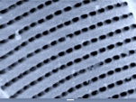
The species 6 and 7 belong to type B.
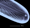
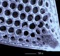
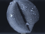
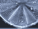
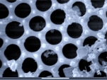
Broken shells display the inner structure very clearly (Fig.8).
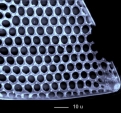
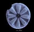
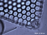
Species of the genus Triceratium (Fig. 9 to 14) show the thin inner wall (type B). This wall can be seen even with a microscope.
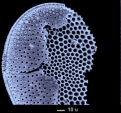
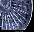
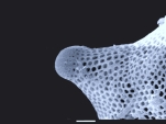
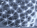
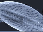
The genus Arachnoidiscus (Fig.15 and 16) provides an unusual image in the SEM: When investigated with a microscope the coarse radial ribs are predominant, but using the SEM you can see them only when the inner surface is inspected (fig.15) - investigated from outside the resulting image is very surprising.
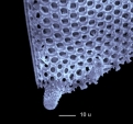
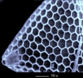
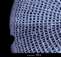
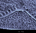
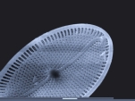
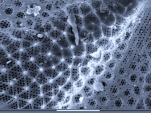
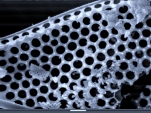
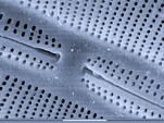
Actinoptychus (the "folded
star", Fig.17 and 18) is the natural enemy of the micro-photographer, because
the deeply corrugated frustules cannot be brought into focus satisfactorily
even when moderately magnified. Using a common microscope one seems to see two
different coarse pore systems masking each other. The true conditions are shown
in figure 18: There is only one single pore system with smaller pores located
in larger ones - assigning them to the shell types defined above is impossible.
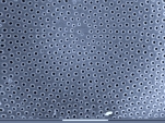
The interior of Epithemia corresponds to the optical image, because the coarse septa can be seen from the outside and the inside as well, they are predominant when inspected with a microscope, but the subtle pore structure is nearly invisible. In particular we cannot detect the intricately shaped apertures (pores which are closed by a leaf-shaped structure).
 |
 |
 |
 |
 |
|
| 21 | 22 | 23 | 24 Biddulphia | 25 Biddulphia | |
 |
 |
 |
 |
 |
|
| 26 Mastogloia | 27 Mastogloia | 28 Mastogloia | 29 Biddulphia | 30 Biddulphia | |
 |
 |
 |
 |
 |
|
| 31 Ardissonia | 32 Cocconeis | 33 Cocconeis | 34 Cocconeis | 35 Actinoptychus | |
 |
 |
 |
 |
 |
|
| 36 Actinoptychus | 37 Actinoptychus | 38 Triceratium | 39 Triceratium | 40 Cymbella | |
 |
 |
 |
 |
 |
|
| 41 Cymbella | 42 Navicula | 43 Navicula | 44 | 45 |
High resolution fotos of diatoms
Identification
keys to the genera according to HUSTEDT ![]()
More pictures of diatoms (Centrales)
More Pictures of diatoms (Pennales)
Copyright: webmaster@mikrohamburg.de