.
.
.
.
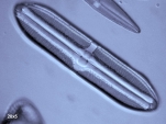
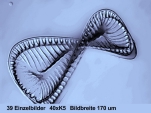
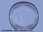
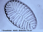
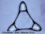
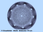
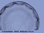
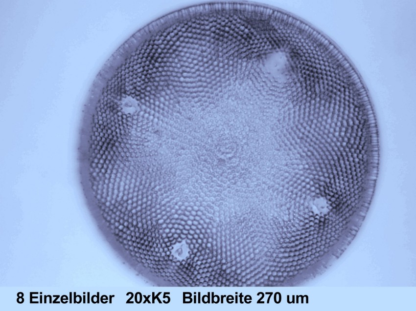
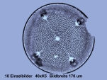
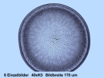
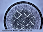
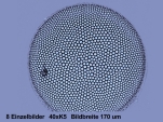
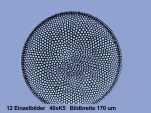
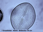
Until recent days making high resolution micrographs of diatoms was a laborious science. Because of the low contrast special sensitive material had to be used and special methods of developing had to be applied. Very often micrographs were insufficient because of the limited depth of field of the objectives and even good micrographs could never display the entire frustule in focus. Today digital cameras in combination with digital image processing have changed the scene completely: You simply make some 5 photoes moving the stage by 1 um, afterwards you select the best image.
|
|
|
|
|
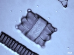
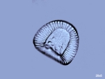
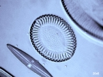
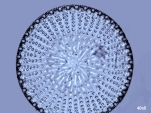
| 1 Biddulphia . |
2 Triceratium | 3 Stictodiscus | 4 Aulacodiscus | 5 Campylodiscus |
|
|
|
|
|
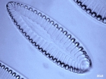
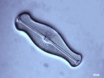
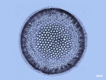
| 6 Surirella . |
7 Surirella | 8 Surirella | 9 Surirella | 10 Didymosphenia |
|
|
|
|
|
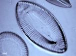
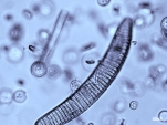
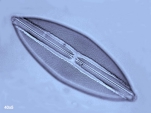
| 11 Epithemia . |
12 Navicula | 13 Pinnularia | 14 Pinnularia | 15 Pinnularia |
|
|
|
|
|
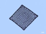
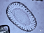
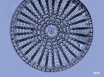
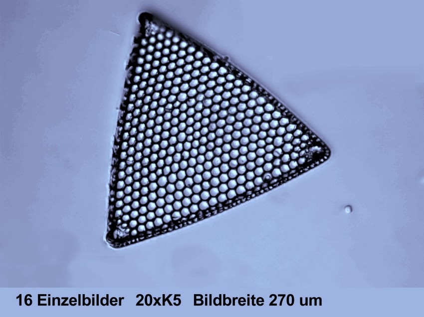
| 16 Stictodiscus . |
17 | 18 Arachnoidiscus | 19 Arachnoidiscus | 20 |
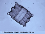
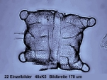
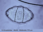
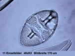
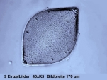
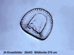
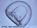
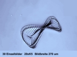
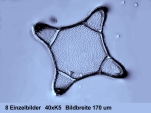
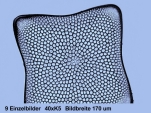
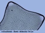
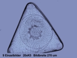
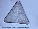
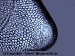
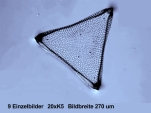
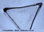
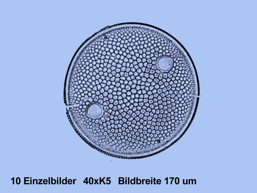
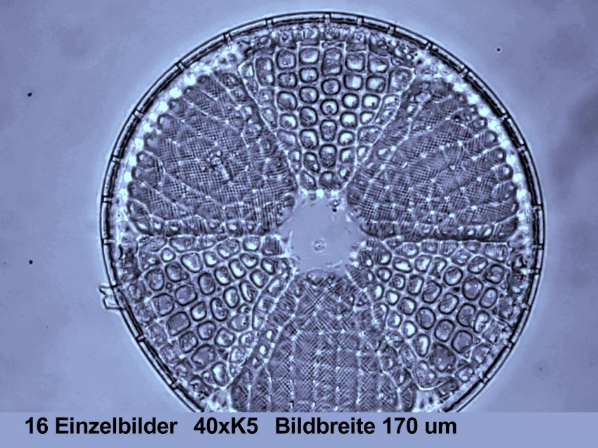
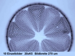
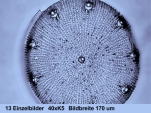
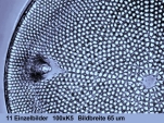
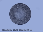
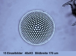
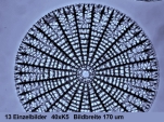
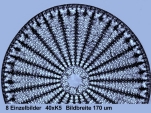
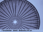
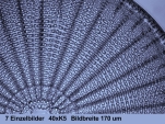
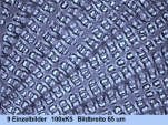
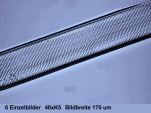
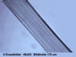
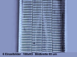
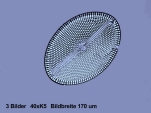
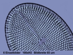
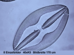
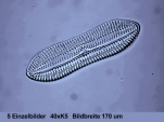
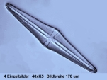
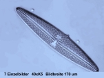
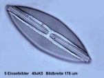
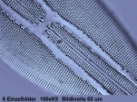
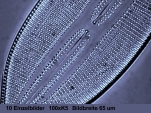
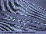
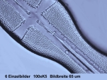
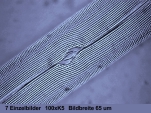
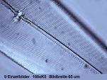
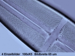
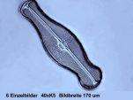
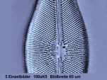
Horizontal width: 20x5: 270 um 40x5: 170 um 100x5: 65 um
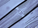
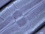
A great advantage are special programs, like HELICON-FOCUS, which is available as share ware. You simply make 10 or 20 single images, moving the stage by 1 um per image, then load this pack of images into the programme and start the processing procedure. HELICON-FOCUS extracts the sharp parts of the images and assembles them to a new image, adding every part in an "intelligent way" to the image just assembled. This procedure requires images with overlapping sharp parts, so the stage must be moved by only 1 um per image. The assembling can be tracked on the screen, as every step needs about one second. So artefacts or ruptures can be detected. In such a case some images must be erased and the whole process must be started again. The proramme can be handled easily, all features are self-explaining.
 |
 |
 |
 |
 |
| 1 Biddulphia . |
2 Biddulphia | 3 Biddulphia | 4 Kittonia | 5 Biddulphia |
 |
 |
 |
 |
 |
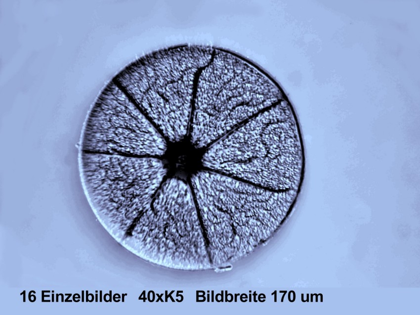
| 6 Campylodiscus . |
7 Campylodiscus | 8 Campylodiscus | 9 Campylodiscus | 10 Campylodiscus |
 |
 |
 |
 |
 |
| 11 Surirella . |
12 Trinacria | 13 Trinacria | 14 Stictodiscus | 15 Stictodiscus |
 |
 |
 |
 |
 |
| 16 Triceratium . |
17 Triceratium | 18 Triceratium | 19 Trinacria | 20 Trinacria |
 |
 |
 |
 |
 |
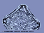
| 21 . |
22 Triceratium | 23 Gyrodiscus | 24 Auliscus | 25 Auliscus |
 |
 |
 |
 |
 |
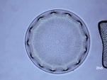
| 26 Actinoptychus . |
27 Actinoptyhus | 28 Aulacodiscus | 29 Aulacodiscus | 30 Aulacodiscus |
 |
 |
 |
 |
 |
| 31 Aulacodiscus . |
32 Aulacodiscus | 33 Aulacodiscus | 34 Coscinodiscus | 35 Coscinodiscus |
 |
 |
 |
 |
 |
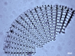
| 36 Arachnoidiscus . |
37 Arachnoidiscus | 38 Arachnoidiscus | 39 Arachnoidiscus | 40 Arachnoidiscus |
 |
 |
 |
 |
 |
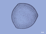
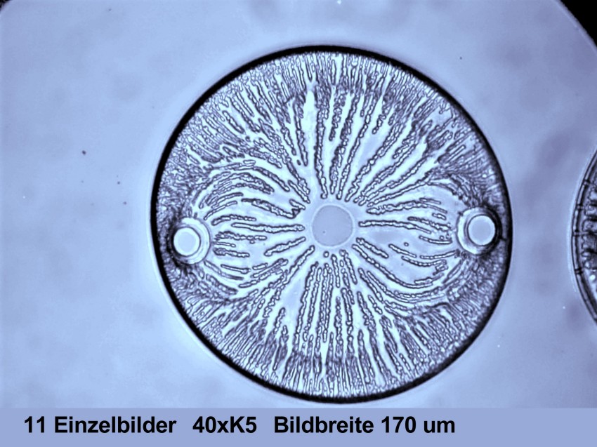
| 41 Actinocyclus . |
42 | 43 | 44 | 45 Ardissonia |
 |
 |
 |
 |
 |
| 46 Ardissonia . |
47 Ardissonia | 48 Mastogloia | 49 Mastogloia | 50 Mastogloia |
 |
 |
 |
 |
 |
| 51
Navicula spectabilis |
52 Diploneis | 53 Navicula | 54 Stauroneis | 55 Trachyneis |
 |
 |
 |
 |
 |
| 56 Navicula . |
57 Navicula | 58 Navicula | 59 Navicula | 60 Stauroneis |
 |
 |
 |
 |
 |
| 61 Gyrosigma . |
62 Pleurosigma | 63 Stauroneis | 64 Didymosphenia | 65 Didymosphenia |
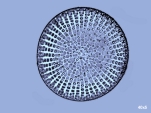
All images on display were taken with a simple digital camera (KODAK EasyShare C613, 6,1 megapixel). The pictures have 2848x2134 pixel corresponding to jpgs of about 0.8 MB. Light source: Low voltage bulb plus interference filter "green". The images have been sampled down to 850x637 pixel.
Copyright: webmaster@mikrohamburg.de