 |
 |
| Squid | Guppy |
To investigate animal tissue, sections must be made of less than 0.01 mm in thickness. The necessary technology is very sophisticated, yet even amateurs can make appropriate preparations, this demonstrate the examples presented here. But usually it is easier to buy microscope slides in shops specialized on such things.
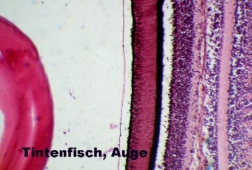
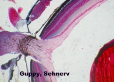
The first two sections show the retina of a squid and the retina of a guppy. You can clearly see the fundamental differences: The retina of the squid is one-layered and the photoreceptors are oriented towards the lens, i.e., the light falls directly on the receptors after having passed the lens . The retina of the guppy is multi-layered and the photoreceptors point away from the lens. So the light must pass all layers of the retina before it reaches the receptors. Besides, the section on display is a rarity, since it shows the "blind spot", that is the very spot where the optic nerve pierces the retina. At this point there are no receptors.
|
|
| Squid | Guppy |
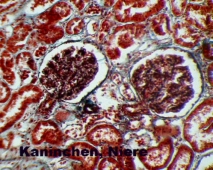
The following section shows the renal cortex of a rabbit. One recognizes two renal corpuscles with their delicate capillaries, each of which are rolled into a ball, the glomerulus. Here the primary urine is pressed from the blood, which then moves into the cavity of Bowman's capsule, and is further concentrated afterwards. The performance of the kidney is evident considering that the human kidneys produce 120 liters of primary urine per day (!) which is then concentrated to about 1 ltr per day.
 |
|
Kidney
|
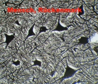
The next picture shows
a section through the human spinal cord. The big pyramid-shaped a-motoneurons
can be seen clearly with their many offshoots, the dendrites. Nerve
cells cannot be stained satisfactorily when using standard methods. Instead
nerve-tissue is impregnated with silver salts, then the sections undergo a
process similar to a black and white photo.
 |
|
Spinal
cord
|
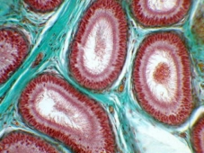
The last picture shows the section through the epididymis of the cat.
 |
|
Epididymis
|
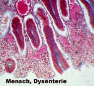
Histological sections
can also demonstrate the damages that are caused by pathogenic microorganisms.
This section shows the wall of the colon of a man who died of tropical
dysentery. The dark-coloured mucous membranes of the krypts are detached
from the surrounding tissue, and towards the intestinal lumen (bottom of the
picture) they seem to have been virtually razed. At the same time we recognize
that the blood vessels of the intestines are filled with blood cells (top
of the picture). Because of such damage the colon is no longer able to reabsorb
water, instead water leaks from the blood vessels into the lumen. The consequence
is severe diarrhoea, accompanied by fluid and salt loss. If untreated, this
leads to a fatal circulatory collapse.
 |
|
Colon,
Dysentery
|