REFLECTED
LIGHT MICROSCOPY
Reflected
light microscopy is a very special part of microscopy, so this method is not
suitable for an amateur: You need special "reflected light microscopes",
and the samples must be absolutely smooth and plane due to the low depth of
field and in order to avoid reflections. Samples must be ground with emery first
to get an even surface, then they are polished carefully and afterwards etched
into the surface to make the structures now blurred by the polishing process
visible again. Only wafers and chips can be analyzed without pre-treatment.
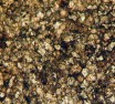
The first picture shows
the etched surface of a metal. One can see that metals consist of many crystals
although to the bare eye they seem to have no structure at all. Along the "grain
boundaries" cracks can easily spread, so investigating surfaces of metals
using reflected light microscopy allowes deductions concerning their mechanical
characteristics. By adding other metals and controlled cooling in alloys defined
textures can be achieved. So this kind of microscopy is a prerequisite both
in material research as well as in material control.
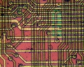
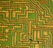
The next images show
the surface of chips. The conducting paths of aluminium seem to be made of gold,
but also the deeper layers appear intensely colored, even though they consist
of colorless materials. All these colors are "interference colors",
which always occur when white light passes through very thin layers - such colors
are well known even in everyday-life: soap bubbles show them, too. In fact the
layers of chips are very thin - they are less than one micrometer thick. A trained
eye can estimate the thickness very accurately by means of the interference
color. So reflected light microscopy is used in chip manufacturing and in process
monitoring.
We would have liked
to demonstrate the structure of modern memory chips, but these structures
are so small that they cannot be resolved adequately with the reflected light
microscope!
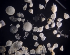
Biological objects
very often produce impressiv images, especially when observed using darkfield
microscopy.
<
HOME >
Copyright: webmaster@mikrohamburg.de